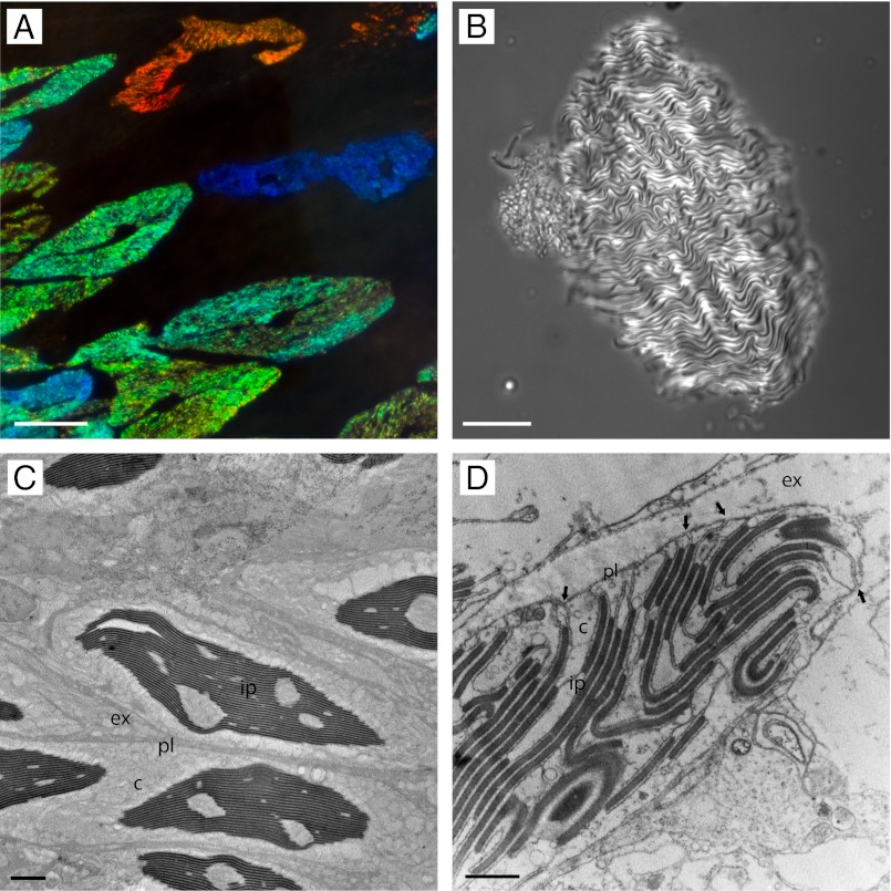
Fig. 1.
Iridophores viewed by light and electron microscopy. (A) Several iridocytes within the dermis (dark-field illumination). (B) A single isolated iridocyte in brightfield illumination. (C) Transmission electron microscopy of several iridocytes in cross-section. (D) A small portion of the edge of an iridocyte, highlighting several key characteristics. The plasma membrane invaginates deep into the cell (arrows) laminating both sides of the cytoplasm-containing platelets and separating them with channels of extracellular space (Scale bars: A, 50 µm; B, 30 µm; C, 3 µm; D, 1 µm.) Ultrastructural features are labeled as c, cytoplasm; ex, extracellular space; ip, iridocyte platelets; pl, plasma membrane.

