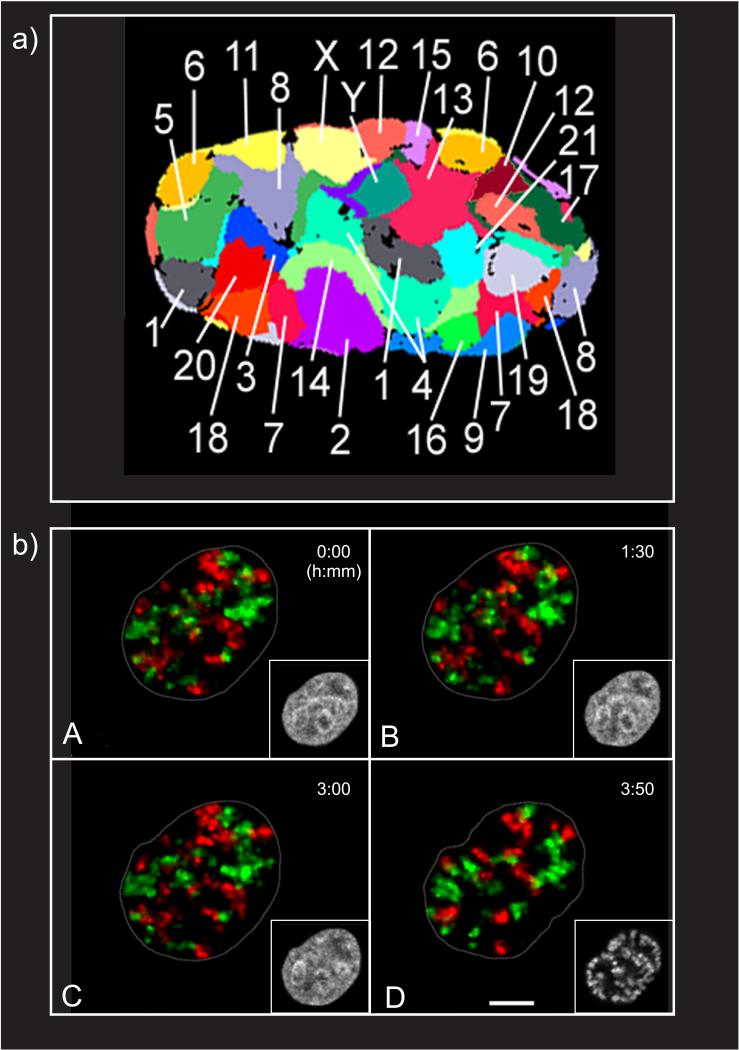
Figure 2.
Chromosomes occupy nonrandom placement in the cell nucleus. (a) Flattened 3D fluorescent in-situ hybridization representation of all 24 chromosome territories (CTs) in a human G0 fibroblast nucleus (166). (b) Stability of CT neighborhood during interphase of living HeLa cells. HeLa cells were replication-labeled during S-phase of two consecutive cell cycles (first cycle, Cy3-dTUP, red; second cycle, Cy5-dUTP, green). (A) Cells were allowed to complete another two cycles before observation was started. (D) After 3 h, 50 min, the cell entered prophase. Frames of maximum intensity projections from light optical serial sections are displayed for the indicated time point. Insets show H2B-GFP signals representing the chromatin (gray) in confocal nuclear midsections. Images are corrected for translational and rotational nuclear movements. Prophase chromosome condensation is a locally confined process with little change to CT arrangements (compare late G2 in subpanel C with early prophase in subpanel D). Scale bar: 5 μm (167). Abbreviations: Cy, cyanine; dUTP, 2′-deoxyuridine 5′-triphosphate; GFP, green fluorescent protein. Adapted from References 166 and 167 with permission.

