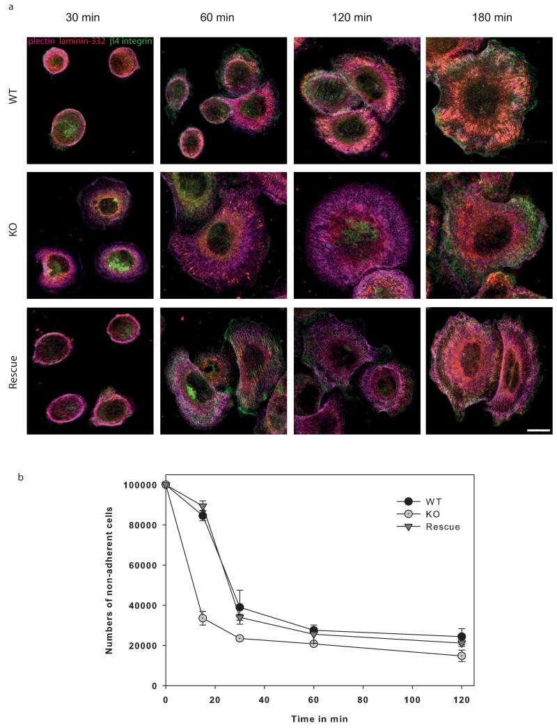
Figure 4. Keratin-free cells adhere much faster.
Immunofluorescence analysis of wildtype and keratin-free keratinocytes in the course of adhesion process after 30 min, 1h, 2h and 3h are shown. Therefore, cells were immunolabeled for hemidesmosomal proteins plectin, β4-integrin, the extracellular ligand laminin-332 and keratin 5. (a) Hemidesmosomal structures visualized by plectin and β4-integrin can be seen after 3h in wildtype keratinocytes. In contrast, keratin-free cells are already adherent after 1h. Moreover, plectin and β4-integrin co-localization is missing in keratin-free keratinocytes. (b) Quantitative analysis of non-adherent cells on specific time points. Bar, 10 μm.

