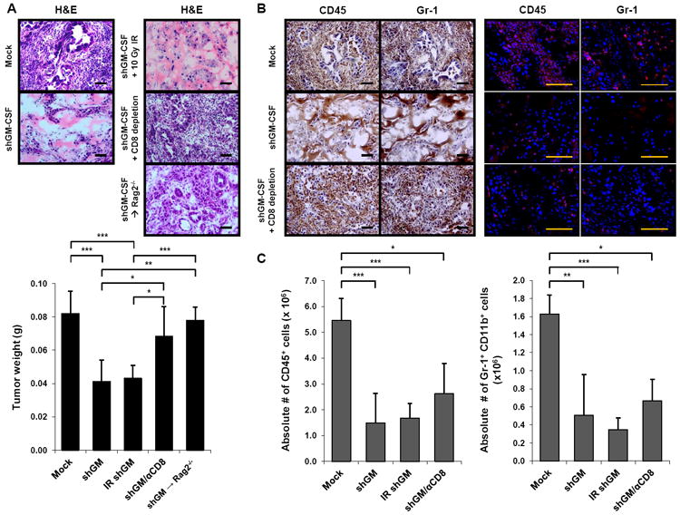
Figure 7. Genetic knockdown of GM-CSF demonstrates that tumor-derived GM-CSF regulates tumor growth and recruitment of Gr-1+ CD11b+ cells in vivo.

(A) Normal control mice or Rag2-/- mice (6-14 weeks) were implanted subcutaneously with either shGM-CSF PDA-1 cells (shGM) or vector only PDA-1 cells (Mock) in matrigel with or without tumor cell irradiation (IR), and with or without intraperitoneal administration of a depleting CD8 mAb. On day 6, tumor growth was evaluated by H&E (top; images of one representative mouse per group are shown) and tumor weight (bottom; data are mean ± SD). n = 3 to 7 mice per group. Scale bars, 50 μM. * indicates p < 0.05, ** p < 0.01, *** p < 0.001.
(B) The immune infiltrate was evaluated by immunohistochemistry (left) and immunofluorescence (right) for CD45 and Gr-1. Images of one representative mouse per group are shown (3 to 7 mice per group). Scale bars, 50 μM.
(C) Absolute numbers of CD45+ cells (left) and Gr-1+ CD11b+ cells (right) were quantified by flow cytometry. Data are mean ± SD. * indicates p < 0.05, ** p < 0.01, *** p < 0.001.
See also Figure S5.
