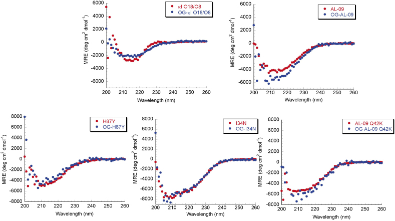
Figure 1. Far UV CD spectra of OG labeled AL proteins show native β-sheet structure.
Data is shown as mean residue ellipticity (MRE) as a function of wavelength. Proteins display the minimum at around 218 nm, characteristic of β-sheets. Experiments were done with 20 μM protein concentration in 10 mM Tris-HCl pH 7.4 at 4°C.

