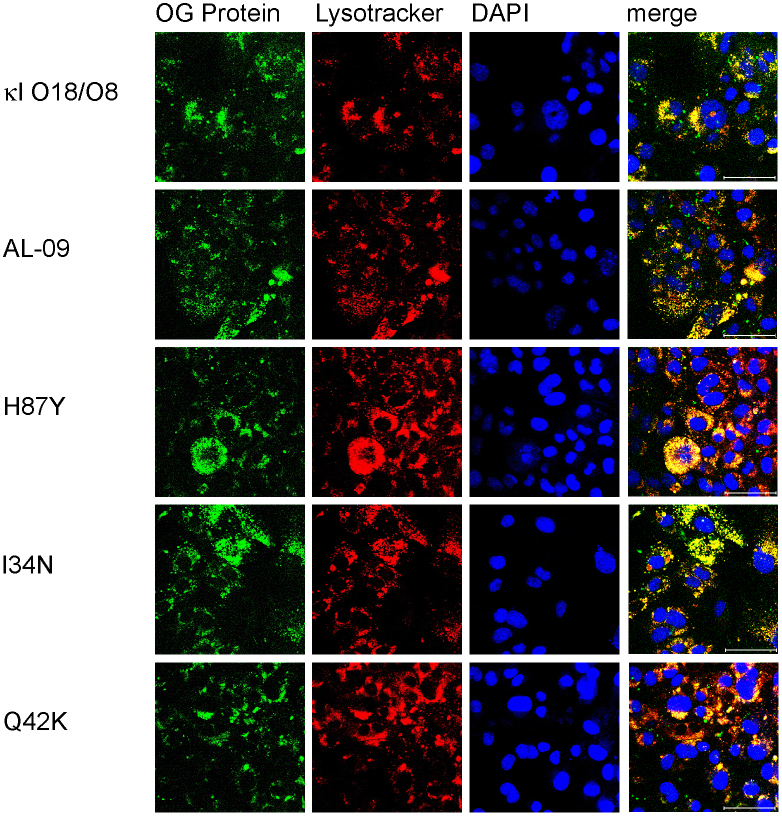
Figure 6. Increased internalization in cardiomyocytes is observed with 1 μM OG-labeled light chain proteins after 72 hours of incubation.
Cardiomyocytes incubated in the presence of different light chains were imaged using confocal microscopy. Oregon green labeled protein (green), lysotracker (red), DAPI (blue), and merged images. The internalization displayed by each protein is stronger than at previous time points. Scale bar is 50 μm.

