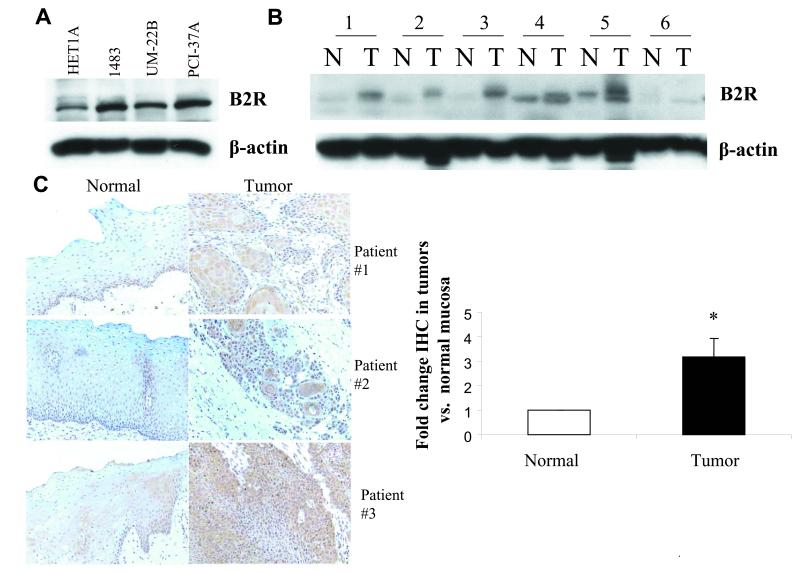
Figure 6. B2 receptor is overpressed in head and neck cancer.
(A) HNSCC cell lines. Cell lysates from an immortalized normal mucosa cell line HET1A and three HNSCC cell lines 1483, UM-22B and PCI-37A were subjected to immunoblotblot analysis to determine B2R levels, and then reprobed with beta-actin. (B) HNSCC tumors compared to normal mucosa by immunoblot. Lysates of HNSCC tumors and corresponding control normal mucosa obtained from six patients were probed for B2 receptor levels by immunoblotting and reprobed with beta-actin antibody. (C) Immunohistochemistry (IHC) of B2R expression in tumor tissue compared to normal mucosa from same patient. IHC staining of B2 receptor was performed in tumor tissue and control normal mucosa from 43 HNSCC patients (Tissue sections from three representative patients). The staining intensity was scored as described in ‘Materials and Methods’ and p-value was computed using the Wilcoxon matched pairs sign rank test.

