Figure 4. .
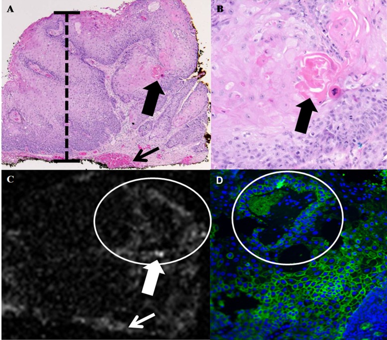
Conjunctival squamous cell carcinoma. (A) Photomicrograph, hemotoxylin and eosin, ×40. The conjunctival epithelium displays faulty epithelial maturation sequencing extending to full thickness (black bar). Invasion of atypical epithelial cells with foci of dyskeratosis is present (large black arrow). A focus of hemorrhage is present at the deep margin of the specimen (small black arrow). (B) Photomicrograph, hemotoxylin and eosin, ×400. Higher magnification view of area denoted by large black arrow in (A) discloses formation of a keratin pearl by atypical epithelial cells within the substantia propria. (C) OCT. Area of atypical tumor cells depicted in (B) is detectable as a focus of increased OCT signal scattering (large white arrow) in (C). Focus of hemorrhage depicted in (A) is also visible as an increase in OCT signal scatter (small white arrow) in (C). (D) Immunofluourescence, ×400. Atypical epithelial cells surrounding the keratin pearl depicted in (B) and visible as increased signal scatter in (C) stained intensely positive on immunofluorescence, indicating a high level of overexpresion of Glut-1 and strong binding by functionalized GNRs.

