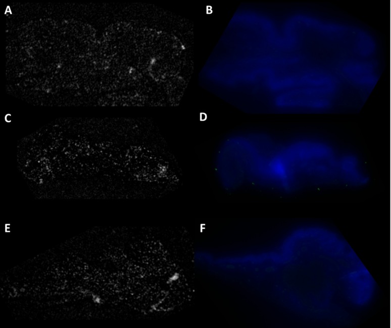
Figure 5. .
OCT images: (A) normal conjunctiva, (C) conjunctival intraepithelial neoplasia: carcinoma in situ (E) conjunctival squamous cell carcinoma. No regions of enhanced scattering were observed, which rules out any nonspecific binding of PAA coated GNRs to tissue. Fluorescence imaging: (B) normal conjunctiva, (D) conjunctival intraepithelial neoplasia: carcinoma in situ (F) conjunctival squamous cell carcinoma. DAPI stains cell nuclei of epithelial cells in blue. No areas of green fluorescence were observed, which rules out the nonspecific binding of Alexa-488 tagged secondary antibody to the tissue surface.

