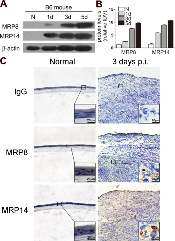
Figure 2. .

Expression of MRP8 and MRP14 in mouse corneas. (A) MRP8 and MRP14 protein levels in B6 corneas were examined using Western blot before and after PA infection. Equal quantities of protein (5 μg) were loaded in each lane. The band intensity of MRP8 and MRP14 (B) was quantitated and normalized to β-actin. Data shown represent one of three individual experiments, each using 5 pooled corneas/time. (C) MRP8 and MRP14 protein expression also was determined by using immunohistochemistry in normal uninfected and infected B6 corneas at 3 days p.i. Magnifications: ×40 (low magnification), ×100 (large inset).
