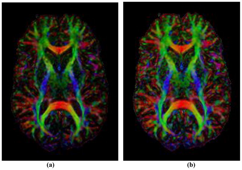
Figure 9. In vivo Color Maps of the Principal Direction (PD) of Tensors in a Human Subject.

(a) Color map based on FA; (b) Color map based on EAR. Compared with the map using FA values, the color map of principal directions is enhanced when using EAR values. Anatomical details are clearer and more discernible. Color schema: red for PDs oriented in horizontal directions; green for vertical; and blue for perpendicular to the viewing plan.
