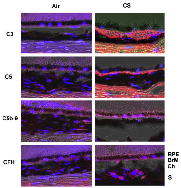
Fig. 5.
Immunolocalization of complement pathway components in mice exposed to chronic cigarette smoke. These confocal immunofluorescence images were overlaid with the bright field images. (Top left panel) Immunoreactivity to C3a is observed in the area of Bruch’s membrane of mice exposed to chronic cigarette smoke. (Second left panel) Immunoreactivity to C5 is observed in the area of Bruch’s membrane of mice exposed to chronic cigarette smoke. (Third left panel) Immunoreactivity to C5b-9 is observed in the area of Bruch’s membrane of mice exposed to chronic cigarette smoke. (Bottom left panel) Immunoreactivity to CFH is observed in the area of Bruch’s membrane of mice exposed to chronic cigarette smoke. (Right panels) Immunoreactivity to C3a, C5, C5b-9 and CFH is not seen in mice raised in air. Scale bar = 20 mm. Blue: DAPI.

