
Figure 1. Location of Human Hypothalamus in vivo MRI and Paraventricular and Supraoptic Nuclei by Histology.
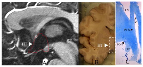
The location of the human hypothalamus (HT) is shown in a mid-sagittal T1 weighted MRI section within the area surrounded by the dashed red outline as projected in the third ventricle (A) and in a coronal section of a grossly dissected human hemisphere within the area surrounded by the dashed white rectangle (B). In C, the paraventricular nucleus (PVN) and the supraoptic nucleus (SON) are indicated by black arrowheads in a coronal histological section stained with thionin. OT = optic tract, LV = lateral ventricle, 3V = third ventricle.