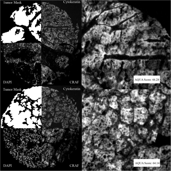
Figure 1.
Example of Automated Quantitative Analysis (AQUA) staining for CRAF in matched primary (upper panels) and metastatic (lower panels) specimens from a single patient: We used anti-cytokeratin antibodies to create a cytoplasmic compartment (two upper right quadrants). A tumor mask was made by filling in holes (upper left quadrants). 4’, 6-diamidino-2-phenylindole (DAPI) defines the nuclear compartment within the tumor mask (left lower quadrants). CRAF expression is measured within the cytoplasmic compartments, within the tumor mask (lower right quadrants), and each clinical case is assigned a score based on pixel intensity per unit area within the tumor mask. The upper panel shows an example of a histospot from a primary specimen and the lower shows the corresponding metastatic tumor. CRAF staining was strong and similar in both specimens.

