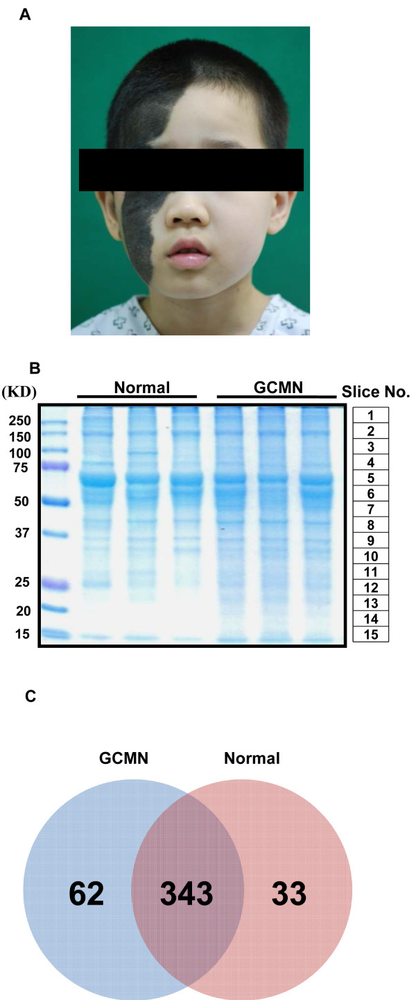
Figure 1.
Comparative proteomic analysis of giant congenital melanocytic nevi (GCMN) and normal skin samples using one-dimensional-liquid chromatography-electrospray ionization-tandem mass spectrometry. (A) A representative GCMN lesion of a donor patient (B) Coomassie-stained gel of normal skin and GCMN proteins (n = 3 each). Slice number indicates the matching mass peak result in Additional file 1: Figure S1. (C) Venn diagram of the identified proteins in normal and GCMN skin samples.

