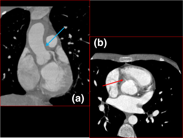
Figure 2.
Coronal (a) and Axial (b) Cardiac Gated CT Chest. Fifty percent narrowing of the origin of the left coronary artery is demonstrated by arrow in (a, blue arrow). The right coronary artery should also be visible in this coronal section but was not due to stenosis (a). It can be seen as a wisp coming off the ascending aorta in the axial image (b, red arrow).

