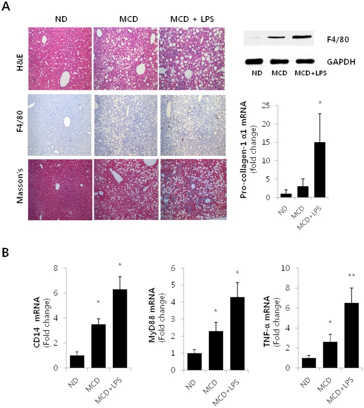
Figure 1. Hepatic Steatosis in LPS-injected MCD diet mice.
(A) Top panels on the left show histology of liver sections from a ND, MCD and LPS-injected MCD mice by Hematoxylin-eosin (H&E) staining (original magnification 200×). Macrophage infiltration was determined by immunohistochemistry analysis of F4/80 (medium panel), and progressive fibrosis was detected by Masson’s trichrome (Masson’s) stain (bottom panel). The right panels, Western blot (top) and qRT-PCR (bottom) analyses were used to quantitate the expression of hepatic F4/80 and hepatic mRNA levels for col1α1, respectively. For the densitometric analysis, n = 3; * P<0.05. (B) Hepatic levels of CD14, Myd88 and TNF-α mRNA were measured by qRT-PCR in ND, MCD and LPS-injected MCD mice. Genes were normalized to18S rRNA as an internal standard and data are shown as fold increase. *P<0.05, **P<0.01.

