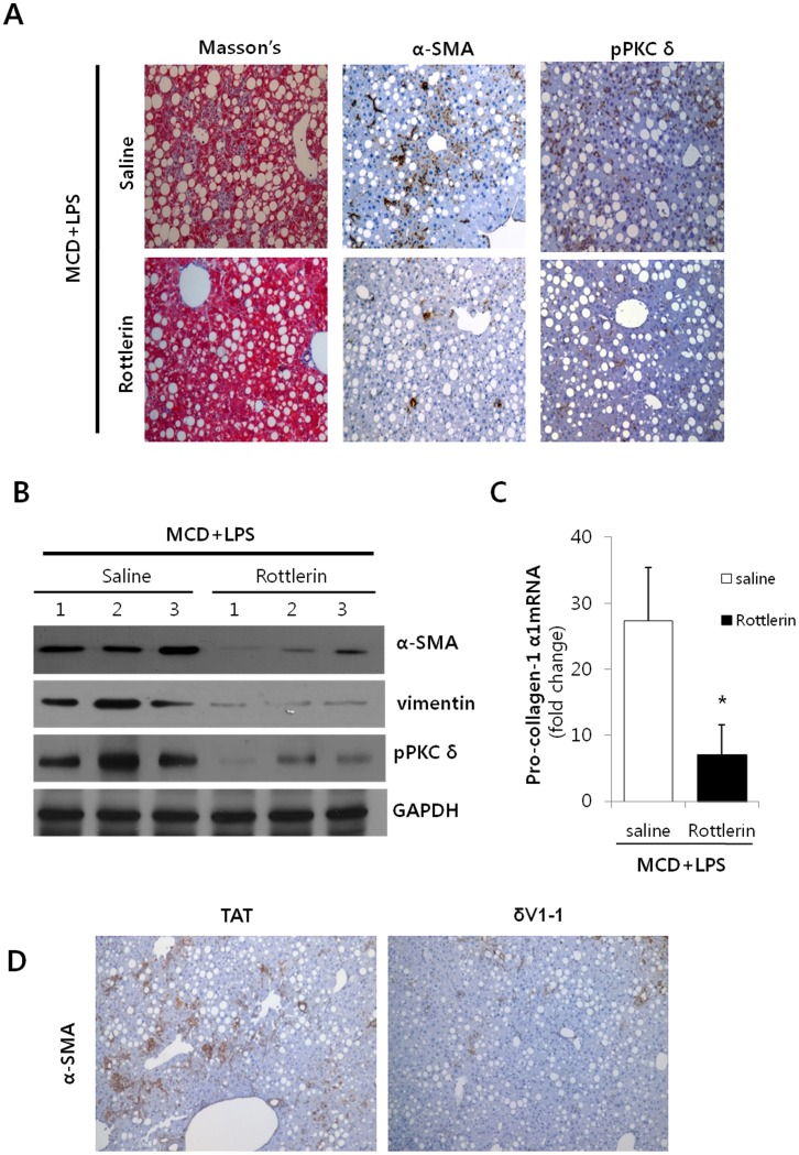
Figure 4. Effects of Rottlerin on LPS-injected MCD mice.
(A) Representative liver tissue sections from LPS-treated MCD mice injected with either saline or Rottlerin (10 mg/kg) were stained with Masson’s trichrome (Masson’s) and α-SMA and phospho-PKCδ antibody (original magnification 200×). (B) Liver tissue extracts were prepared from mice fed the MCD diet for 3 weeks and subsequently injected with 2.5 mg/kg LPS and 10 mg/kg Rottlerin. α-SMA, vimentin and phospho-PKCδ protein content were quantified by Western blot using equal amounts of total liver proteins. The levels of α-SMA and vimentin were observed to significantly decrease in the Rottlerin treatment group. For statistical significance, three liver extracts from each individual were used for each group. Expression levels were normalized relative to GAPDH. (C) mRNA levels of hepatic collagen were determined by qRT-PCR analysis. Data plots represent the mean ± SD of three independent experiments. *P<0.05 versus saline group. (D) Indicated LPS-injected MCD diet mice were pretreated with TAT or δV1-1 peptide (0.2 mg/kg) for 1 hour. Representative liver tissue sections from LPS-treated MCD mice injected with either TAT or δV1-1 peptide were stained α-SMA with antibody (original magnification 200×).

