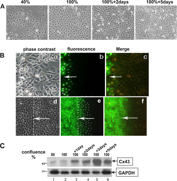
Figure 1.
Cell density increases GJIC and Cx43 levels.A. Immortalised lung epithelial E10 cells were plated in 3 cm plastic petri dishes, grown to different densities and photographed under phase-contrast illumination. Magnification: 240x. B. E10 cells were plated in electroporation chambers and subjected to a pulse in the presence of Lucifer yellow when 90% confluent (a-c) or 3 days after confluence (d-f) and photographed under phase-contrast (a, d), fluorescence (b, e) or combined (c, f) illumination (see Methods, Figure 7). Arrows point to the position of the edge of the electroporated area. In a, b, d and e, stars mark cells loaded with the dye at the edge of the electroporated area and dots mark cells into which the dye was transferred through gap junctions. Magnification: 240x C. E10 cells were seeded in plastic petri dishes and when they reached the indicated densities, detergent cell extracts were probed for Cx43 (top) or GAPDH (bottom) as a control.

