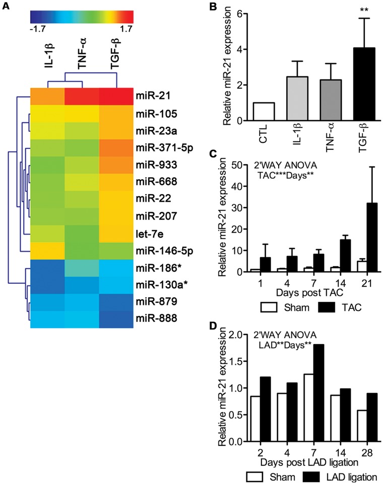
Figure 3. miR-21 is highly up-regulated during fibrogenic EMT.
Differentially expressed miRNAs were assessed in EMC cultures stimulated for 48h with IL-1β, TNF-α, or TGF-β. A, Cluster analysis and heatmap of regulated miRNAs (fold change (FC) above 2 or under −2) as detected by microarrays and compared to non-stimulated control cells. B, Validation of miR-21 expression after 48h of EMT induction in EMCs by qRT-PCR (means+SD, n = 3; standardized to the control (CTL)). Statistical significance was tested by one-way ANOVA and treatment effects by Tukey’s post test. miR-21 up-regulation was further validated by qRT-PCR in vivo using both a model of C, pressure-overload (transverse aortic constriction; TAC) and D, myocardial infarction (ligation of left anterior descending artery; LAD). These data were acquired on biological triplicates (means+SD) or a pool of 1–4 hearts, respectively, and analyzed against sham treated animals by two-way ANOVA. All miRNA qRT-PCR data were normalized against miR-17 and miR-195. **P<0.01, ***P<0.001 vs. control.

