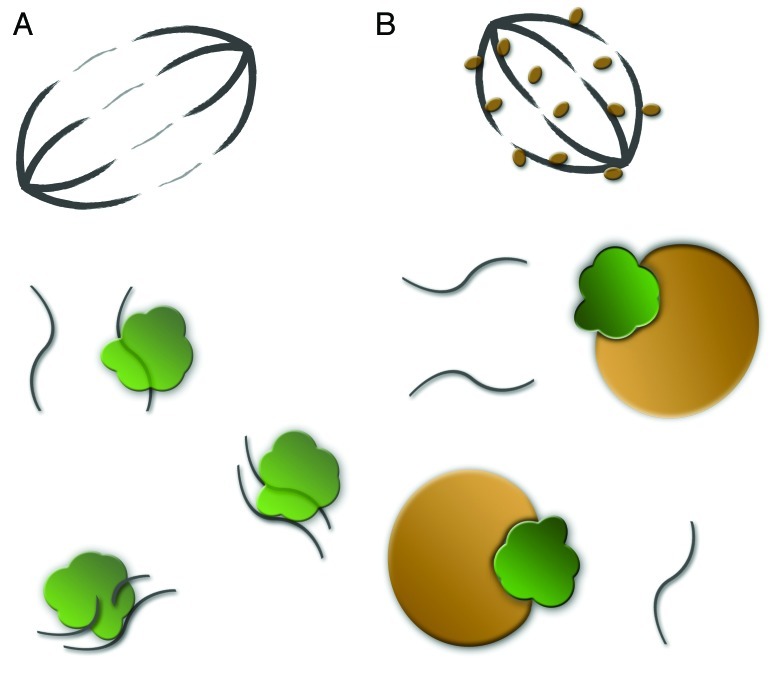
Figure 1. (A) Cartoon of a normal dividing cell with meotic/mitotic spindle at the top. Complexes of Argonaute proteins (green) and antisense TE fragments capture and destroy TE mRNAs. (B) Dividing cell infected with Wolbachia. Wolbachia (brown) associate with microtubuli (top) and capture Argonaute proteins (green). TE derived mRNAs are left to insert back into the host genome.

An official website of the United States government
Here's how you know
Official websites use .gov
A
.gov website belongs to an official
government organization in the United States.
Secure .gov websites use HTTPS
A lock (
) or https:// means you've safely
connected to the .gov website. Share sensitive
information only on official, secure websites.
