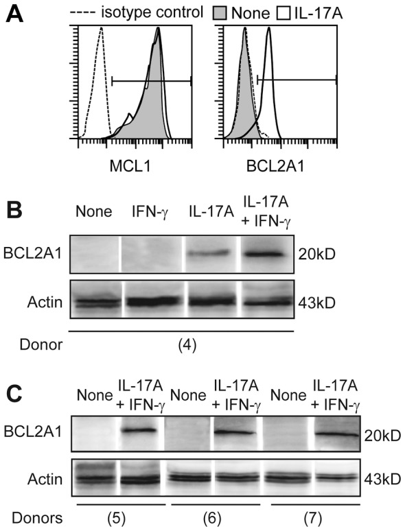
Figure 4. MCL1 and BCL2A1 protein expression in IL-17A-treated DC.

(A) Intracellular expression of MCL1 and BCL2A1 in DC treated (white) or not (gray) with IL-17A, at day 7, representative of n>3, SD<2%. (B,C) Western blot analysis of BCL2A1 versus actin protein expressions in DC cultured with indicated cytokines, lyzed at day 5 for 4 donors (4) to (7), in separated experiments.
