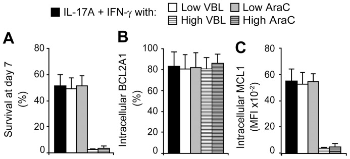
Figure 8. Survival and phenotype of DC cultured with VBL or AraC.

DC were stimulated with IL-17A and IFN-γ for 7 days. According to their toxicity kinetic, VBL or AraC were added in the culture at day 6 or 5, respectively. (A) Cell survival assessed by DiOC6 and PI staining, at day 7. (B) BCL2A1 and (C) MCL1 intracellular expressions were measured prior DiOC6 and PI staining. “Low” doses of VBL and AraC were 0.06 and 4 µM, respectively. “High” doses were ten times more. Mean and SD of n = 5.
