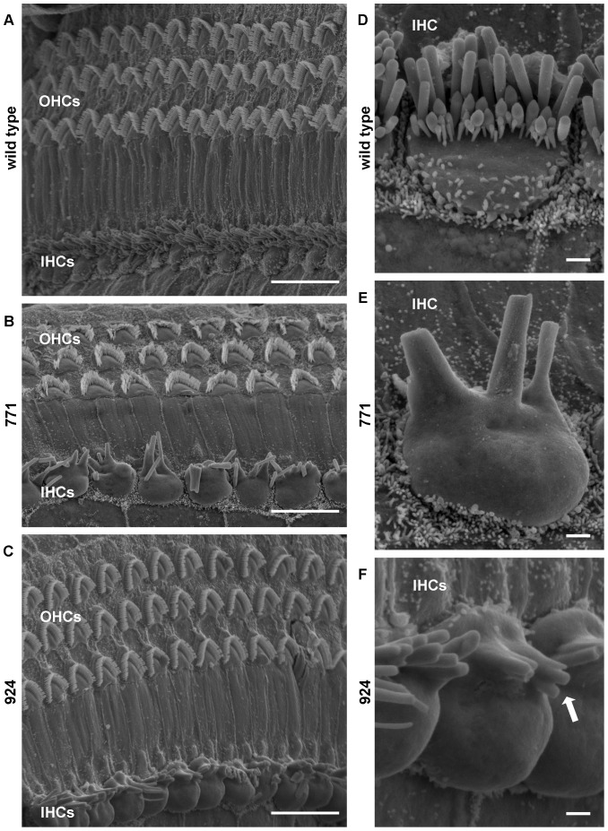
Figure 5. IHC stereocilia abnormalities in Diap3-overexpressing mice at 24 weeks of age.
Scanning electron microscopy images of the organ of Corti were obtained from wild-type mice, line 771, and line 924. Scale bars in A–C represent 10 µm; scale bars in D–F represent 1 µm. (A) Stereocilia of wild-type OHCs and IHCs appeared distinct and organized. (B–C) In contrast, IHC stereocilia of line 771 (B) and line 924 (C) display abnormal fusion, occur in a single row, and are substantially reduced in numbers. In some regions, OHC hair bundles were splayed or flattened (B). At higher magnification, the fused appearance of the IHC stereocilia in line 771 (E) and line 924 (F) becomes more obvious in comparison to the wild-type hair bundle (D). Note the stereocilium marked with an arrow in (F) that appears to have three tips to a single base. Apical bulging of the IHC cell bodies is noted but may represent artifact.

