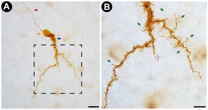
Figure 3. Representative photomicrograph of a Cajal-Retzius cell stained by the new photooxidation method.
A: Low magnification (40× objective lens) photomicrograph of the DAB-stained Cajal-Retzius Cell obtained from a transverse section of developing rat neocortex. Note that the cell is stained over its entire volume, with no variations in staining intensity. The red arrow points at the cell's axon. The blue arrow indicates the large horizontal dendrite that defines the Cajal-Retzius cell type. The orange arrow points at the cell's soma Scale bar corresponds to 20 µm. B: High magnification (100× objective lens) photomicrograph of the insert indicated in A (dotted line square). Note the successful staining of dendritic shafts and spine-like appendages (green arrows), highlighting the high degree of detail and acuity of the staining. Scale bar corresponds to 10 µm.

