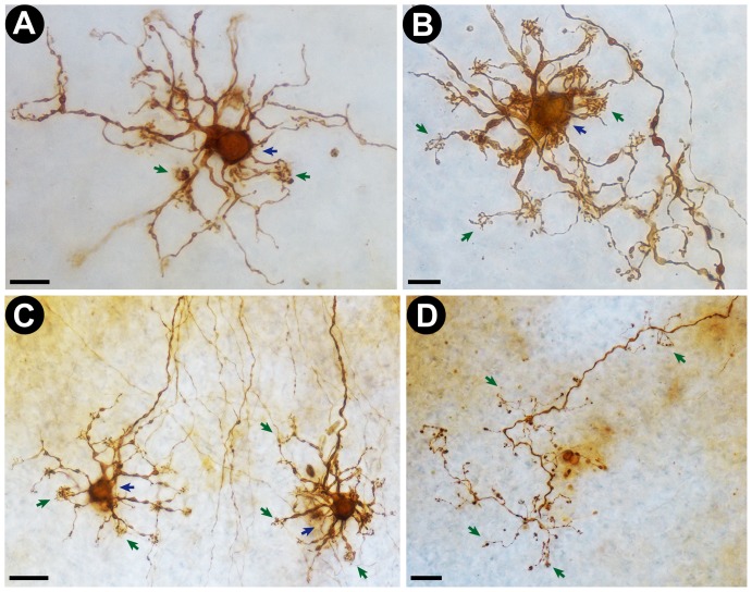
Figure 4. Representative photomicrographs of human retinal neurons and axons stained by the new photooxidation method.
Cells were processed and observed in intact post-mortem human retinas. A and B: High magnification (100× objective lens) photomicrographs of DAB-stained putative horizontal retinal neurons. Note the clear delineation of the cell body (blue arrows) and dendritic tufts (green arrows). Scale bars correspond to 10 µm. C: Low magnification (40× objective lens) photomicrograph of two DAB-stained retinal cells (putatively horizontal cells). Scale bar corresponds to 20 µm. D: Low magnification (40× objective lens) photomicrograph of a DAB stained axon terminal in a sample of human retina. Note that even fine structures, such as axonal knobs (arrows), are clearly stained. Scale bar corresponds to 20 µm.

