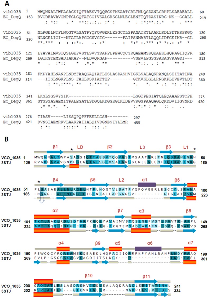
Figure 1. Sequence alignment of VCO395_1035 with E. coli DegQ.
A. Sequence alignment of the query (vib1035) and the E. coli DegQ (EC_DegQ). The ‘*’ indicate the conserved amino acids; ‘:’ represents similar group of amino acids. B. Sequence alignment used for 3D-modeling of VCO395_1035 using E. coli DegQ as template (PDB ID: 3STJ). The blue arrows indicate β sheets, orange bars indicate helix and the yellow bars indicate loops. The deep blue color indicates identical amino acids; lighter blue colors indicate similar and weakly similar amino acids. The two major loop modeled to their corresponding secondary structure were shown in violet color. The predicted catalytic triad residue Ser12-His50-Leu52 indicated by ‘*’ and conserved Ser53 residue with DegQ Ser187 which is one of catalytic triad residue of DegQ of E. coli indicated by down arrow.

