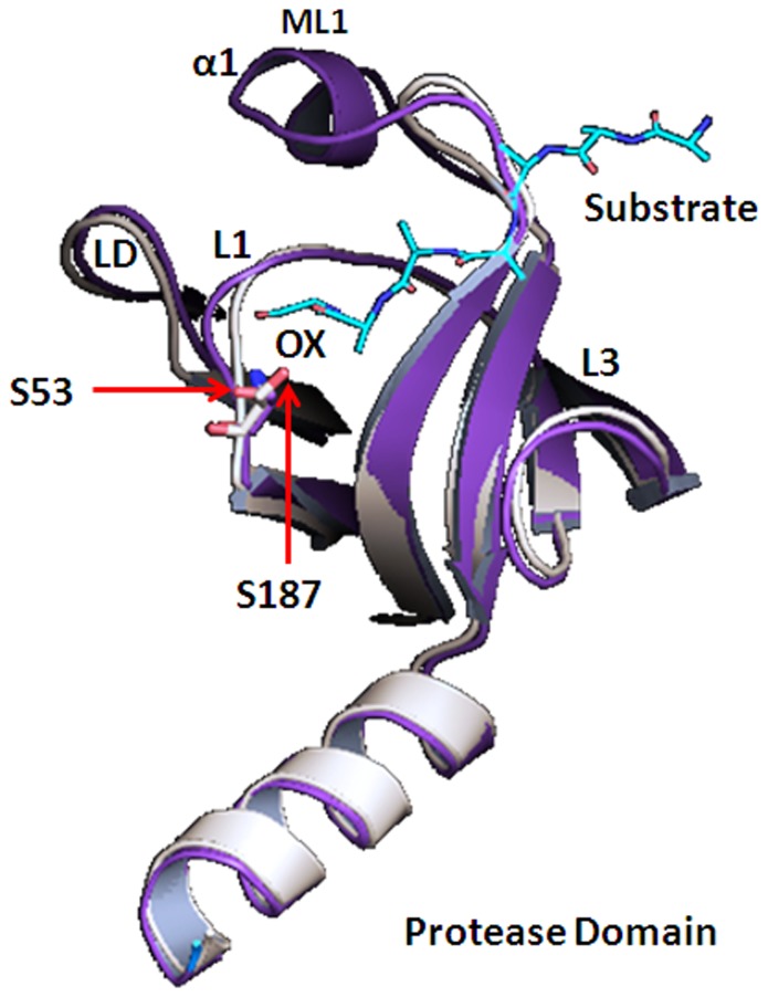
Figure 3. Structural alignment of protease domain.
The cartoon representation of protease domain of model VCO395_1035 (magenta) aligned with template 3STJ (light orange) showing conserved Ser53 with DegQ Ser214 which is one of catalytic triad residue of DegQ along with substrate (cyan) bound to active site in Oxyanion hole(ox).

