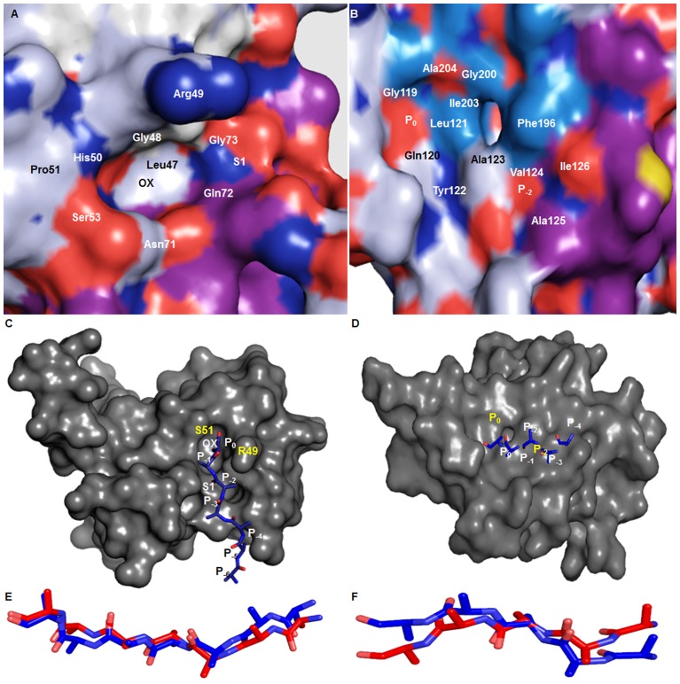
Figure 4. Active site and Protein-substrate interaction using Hex 5.0.
A. The surface view of protease domain containing active site showing the oxyanion hole and properly oriented shallow S1 hydrophobic pocket. B. The surface view of PDZ1 containing hydrophobic binding groove formed by CBL and α7-Helix showing shallow P0 and P−2 substrate binding pocket. C. The C-terminal of poly-alanine peptide substrate (blue) docked into active side of protease domain. D. The C-terminal of poly-alanine peptide substrate (blue) docked into active side of PDZ1 domain via β-aggumentation. E. The superimposition of substrate docked into the protease active site (blue) with respective to template (3STJ) substrate (red). F. The superimposition of substrate docked into the active site PDZ1 domain (blue) with respective to template (3STJ) substrate (red).

