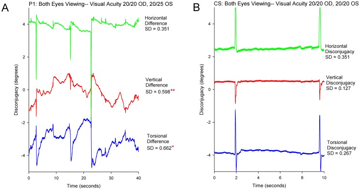
Figure 5. Representative records comparing the disconjugacy of gaze for each directional component for P1 with monocular visual impairment versus a control subject.
(A): Record of P1. (B): Record of control subject. Values shown at right of each plot are SD of the difference between right and left eye position for each directional component. One asterisk indicates that the SD value is significantly larger (p<0.05) than pooled data from normal subjects; two asterisks indicate p<0.01. See caption to Figure 2 for further details.

