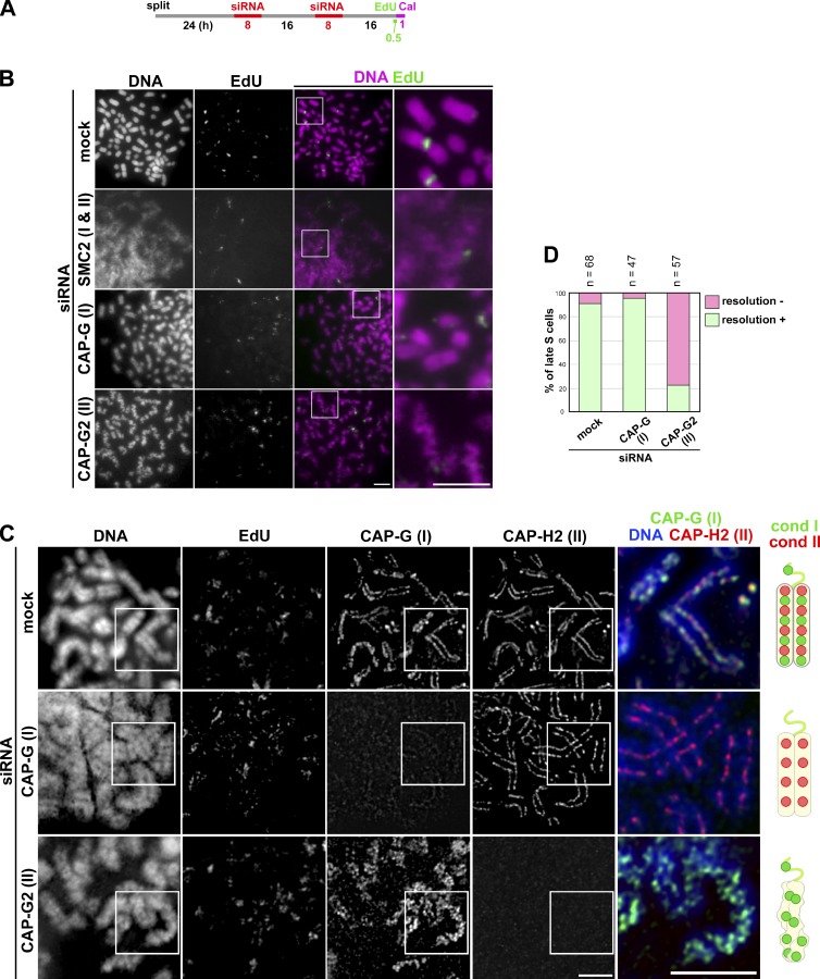
Figure 3.
Condensin II plays a key role in PCC-driven sister chromatid resolution in late S phase cells. (A) Experimental protocol for calyculin A–induced PCC from siRNA-treated HeLa cells. After two rounds of siRNA transfection, the cells were pulse labeled with EdU and then treated with calyculin A (Cal). (B) Cells were processed as in Fig. 2 B. Shown here are representative images of late S-PCC cells from populations mock depleted or depleted of SMC2, CAP-G, or CAP-G2. Bars, 5 µm. (C) HeLa cells (mock depleted or depleted of CAP-G or -G2) were pulse labeled with EdU and treated with calyculin A as described in A. The cells were fixed and immunolabled with antibodies against CAP-G and -H2, as described in Fig. 2 C. Shown here are representative images of late S-PCC cells, along with their merged closeups. Bars, 5 µm. Cartoons depict late S-PCC chromosomes observed under the different conditions. (D) Plotted here are the frequencies of resolution defects in late S-PCC products observed under the three conditions. This quantitative evaluation was completed once. For additional information, see Fig. S5 (A–C).

