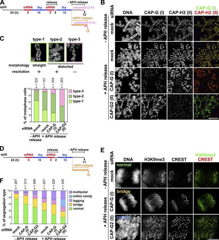
Figure 6.
Application of mild replicative stress to condensin II–depleted cells exacerbates their defects in chromosome architecture and segregation. (A) Experimental protocol for enriching metaphase cells from siRNA-treated synchronized cultures. Cells were harvested at the time points indicated by the arrows after colcemid treatment. (B) Metaphase chromosome spreads were prepared and immunolabeled with antibodies against CAP-G and -H2. Bar, 5 µm. (C) The morphology of metaphase chromosomes observed were classified into three types and plotted for each condition tested. The data shown are from a single representative experiment out of two repeats. For additional information, see Fig. S5 (D and E). (D) Experimental protocol for enriching mitotic cells from siRNA-treated synchronized cultures. No colcemid treatment was applied in this protocol. (E) HeLa cells treated as described in A were fixed on coverslips and immunolabeled with anti-H3K9me3 and a CREST serum. Shown here are representative images of cells displaying anaphase or anaphase-like chromosome morphology: normal segregation, chromosome bridges, and cotton candy chromosomes. Bar, 10 µm. (F) Defects in chromosome segregation were classified into five categories and plotted for each condition. The data shown are from a single representative experiment out of three repeats.

