Figure 8.
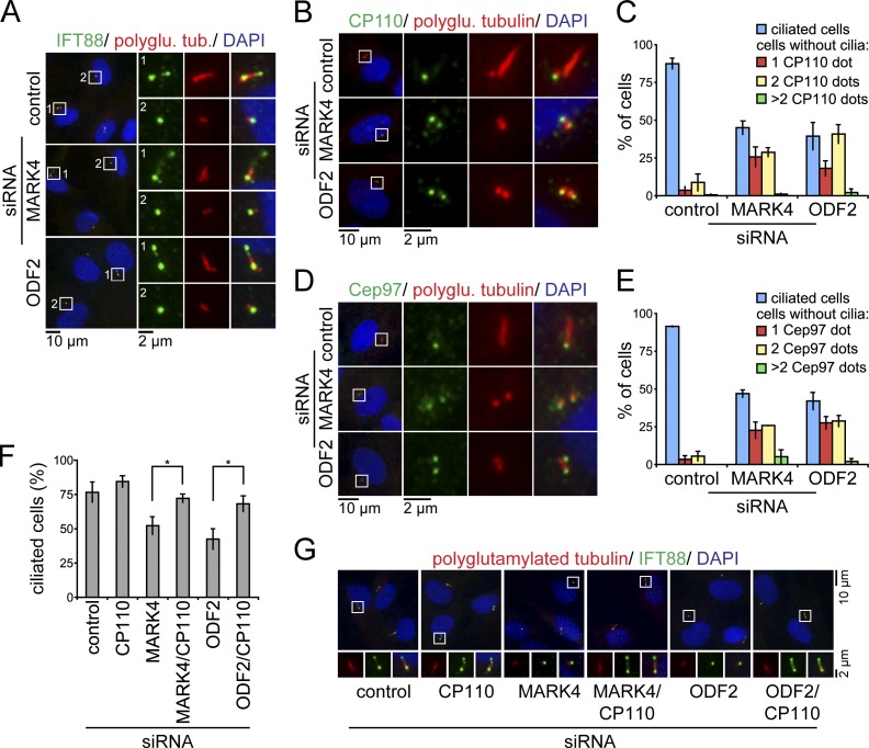
CP110 persists at the mother centriole after MARK4 depletion. (A) RPE1 cells, treated with the indicated siRNAs, were serum starved for 48 h and stained for IFT88, polyglutamylated tubulin (polyglu. tub.), and DNA. (B–E) Same as in A; however, cells were stained for CP110 (B) or Cep97 (D), polyglutamylated tubulin, and DNA. (C and E) Quantification of B and D, respectively. The number of ciliated cells and CP110 (C) or Cep97 dots (E) per centrosome are indicated. (F and G) Codepletion of CP110 rescues the loss of cilia in MARK4- or ODF2-depleted RPE1 cells. (F) After treatment with control, MARK4, or ODF2 siRNA for 24 h, RPE1 cells were subjected to control or CP110 depletion for 24 h and subsequent serum starvation for 24 h. Percentages of ciliated cells based on polyglutamylated tubulin as a cilia marker. *, P < 0.05. (G) RPE1 cells were treated as in F and stained for IFT88, polyglutamylated tubulin, and DNA. In A, B, D, and G, merged images are shown. Regions within the white boxes are shown at higher magnification at the left (A, B, and D) or in the bottom images (G). Data are means ± SD of three independent experiments.

