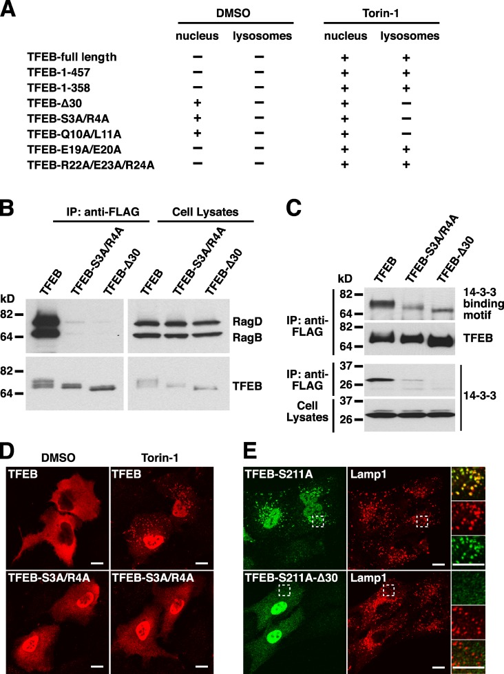
Figure 6.
The N-terminal region of TFEB is necessary for interaction with Rag heterodimers and lysosomal localization. (A) Summary of the nuclear and lysosomal distribution of several TFEB amino acid and deletion mutants in ARPE-19 cells treated with either DMSO or Torin-1. (B) ARPE-19 cells were nucleofected with the indicated Rag- and TFEB-expressing plasmids. After 12 h, cell lysates were immunoprecipitated with the anti-FLAG antibody and analyzed by immunoblotting with antibodies against FLAG and GST (used to detect TFEB and Rag proteins, respectively). (C) FLAG-tagged TFEB-WT or TFEB deletion mutants were immunoprecipitated with the anti-FLAG antibody and analyzed by immunoblotting with antibodies against FLAG, 14-3-3 binding motif, or 14-3-3. (D) Immunofluorescence confocal microscopy showing the subcellular distribution of TFEB-WT and TFEB-S3A/R4A mutant upon incubation with DMSO (vehicle) or 250 nM Torin-1 for 1 h. Cells were fixed, permeabilized with 0.2% Triton X-100, and stained with antibodies against FLAG (used to detect TFEB). (E) ARPE-19 cells expressing either TFEB-S211A or TFEB-S211A-Δ30 were double stained with antibodies against TFEB and Lamp1. Regions within the dotted boxes are magnified in the insets. IP, immunoprecipitation. Bars, 10 µm.

