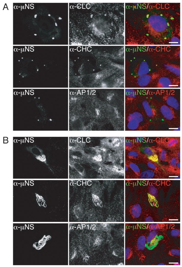
Figure 2. Clathrin LCA/B and clathrin HC, but not AP1 or AP2, colocalize with μNS in both globular and filamentous factory-matrix structures.

A) μNS-WT protein was expressed in CV-1 cells by plasmid-based transfection. At 24 h p.t, cellular localizations of μNS as well as clathrin LCA/B, clathrin HC or AP1/2 were visualized by indirect IF microscopy using a combination of μNS-specific serum antibodies (α-μNS) and MAb CON.1 (α-LC), X22 (α-HC) or 10A (α-AP), respectively, as labeled. B) μNS-WT and μ2 proteins were co-expressed in CV-1 cells by plasmid-based transfection. At 38 h p.t., cellular distributions of indicated proteins were visualized as in A. In A and B, merged images are shown at right (α-μNS, green; α-CLC, α-CHC or α-AP1/2, red). Scale bars, 20 μm.
