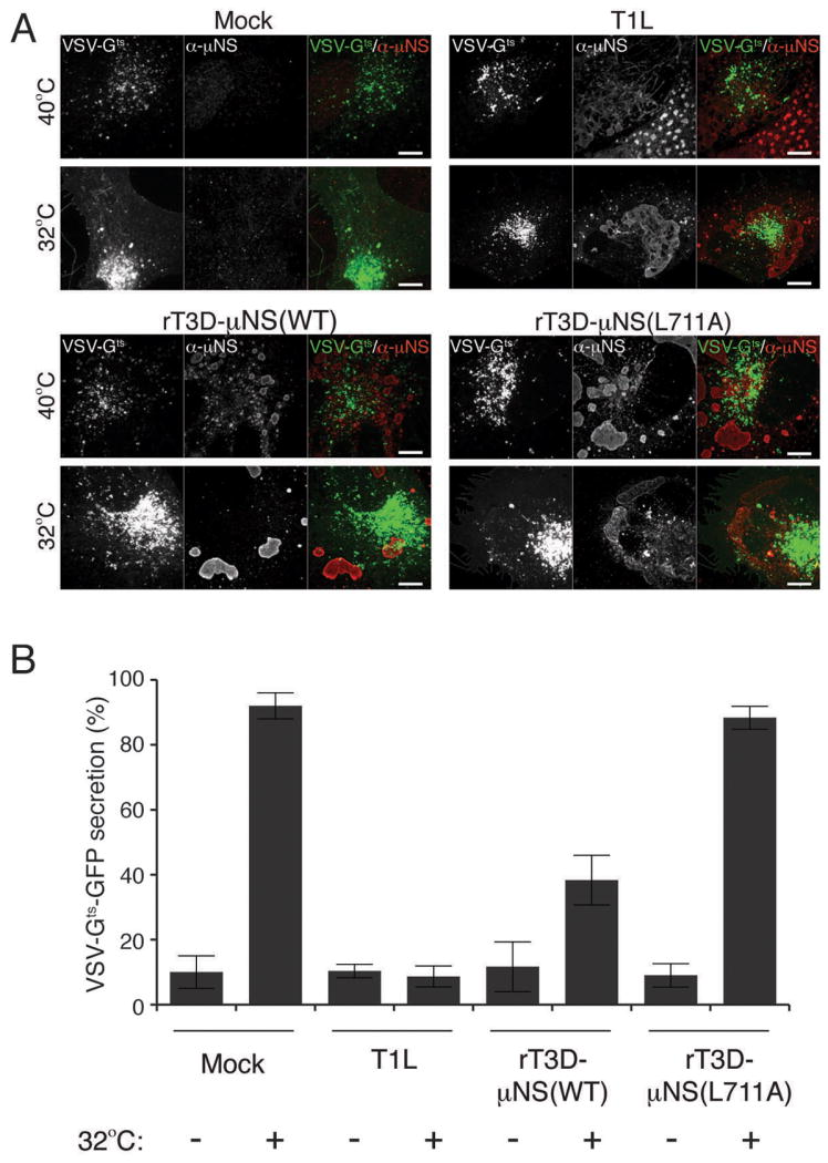
Figure 7. Sequestration of clathrin in MRV factories blocks secretion of VSV-Gts-GFP.

BSC-1 cells were transfected with VSV-Gts fused to GFP and incubated overnight at 40°C. Cells were then mock-infected or infected with either T1L, rT3D-μNS(WT) or rT3D-μNS(L711A) MRV and incubated at 40°C for 12-16 h. 2 h prior to fixation, cells were either kept at 40°C or transferred to 32°C to allow VSV-Gts-GFP protein refolding and secretion from the TGN to the plasma membrane. Fixed cells were immunostained using μNS-specific serum antibodies (α-μNS) and observed by confocal microscopy. A) Representative confocal images of mock- and MRV-infected cells, either kept at 40°C (top panels) or shifted to 32°C (bottom panels). Merged images are shown at right (VSV-Gts-GFP, green; α-μNS, red). Scale bars, 10 μm. B) Bar plot depicting the percentage of cells, which showed plasma-membrane localization of VSV-Gts-GFP in addition to the perinuclear staining after the shift to 32°C (VSV-Gts-GFP-secretion positive). The plot shows the average and the standard deviation of 3 independent experiments, each scoring at least 50 VSV-Gts-GFP-expressing, either mock- or MRV-infeceted (μNS-positive) cells.
