
Figure 1.
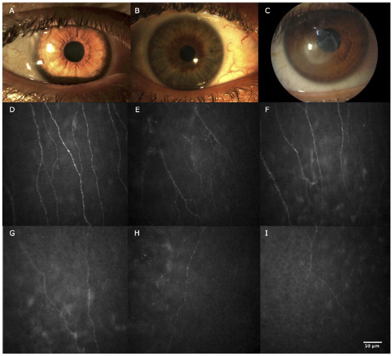
Slit-lamp photographs and in vivo confocal microscopy images obtained at the level of the corneal subbasal nerve plexus. A, Slit-lamp photograph of normal cornea. B, Slit-lamp photograph of contralateral clinically unaffected eye of herpes zoster ophthalmicus (HZO) patient. C, Slit-lamp photograph of eye affected by HZO. D, In vivo confocal microscopy image showing normal corneal subbasal nerve plexus. E, In vivo confocal microscopy image showing contralateral clinically unaffected eye of HZO patient. Note the decrease in length and number of nerves. F, In vivo confocal microscopy image showing HZO with normal corneal sensation. Note the decrease in nerve length. G, In vivo confocal microscopy image showing HZO mild sensation loss. Note the decrease in length and number of nerves and branches. H, I, In vivo confocal microscopy image showing HZO severe sensation loss.