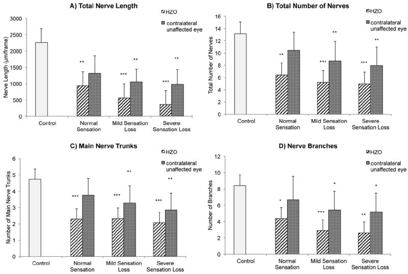
Figure 2.

Bar graphs showing subbasal corneal nerve alterations in affected and clinically unaffected eyes in herpes zoster ophthalmicus (HZO) patients and according to grade of hypoesthesia and in the control group: (A) total nerve length, (B) total number of nerves, (C) main nerve trunks, and (D) nerve branches. Error bars represents standard deviation from the mean. *P<0.05, **P<0.001, and ***P<0.0001 compared with control group by analysis of variance.
