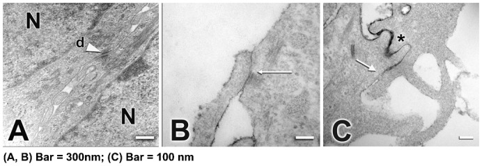
Fig. 1.

Ultrastructure of 4 day (A–C) post-confluent cultured epithelia formed by RCE1(5T5) cells. It is shown the presence of desmosomes (d) between cells (A, arrowhead). TJ were observed at the upper layers of the epithelial sheet, mainly in 4-day confluent epithelia as small direct contact sites between the plasma membranes of two adjacent cells (B, arrows). (C) Note that diffusion of ruthenium red throughout the intercellular space (asterisk) was stopped at TJ location (C, arrow). N, nucleus. In A,B, Bar = 300 nm; in C, Bar = 100 nm.
