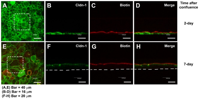
Fig. 2. Paracellular permeability of the cultured corneal epithelia was examined by use of the low molecular weight tracer, EZ-Link sulfo-NHS-LC-biotin.

The tracer did not penetrate the epithelial sheet neither 1 day after confluence (7 days in cell culture) (A–D), nor in 7 day confluent epithelia (E–H), as indicated by the (C,G) lack of staining of biotinylated proteins at cell–cell boundaries in the lower layers of the epithelium. (D,H) Maximal projections of the merged channels, transverse optical sections of cultures stained for cldn-1 to immunolocalize TJ (green channel; B,D,F,H). Proteins biotinylated with the tracer are stained in red (C,D,G,H). (A,E) show the aspect of cldn-1 in xyz maximal projections of the stained cell cultures. Dashed squares show the fields examined in the transverse sections. Dashed lines indicate the basal side of the cultured epithelia. In A,E, Bar = 40 µm; in B–D, Bar = 16 µm; in F–H, Bar = 20 µm.
