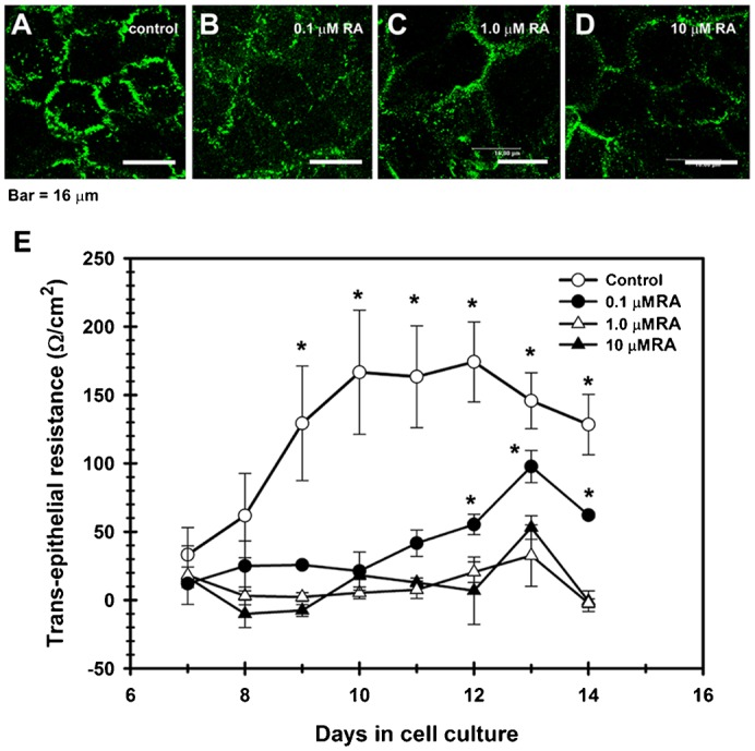
Fig. 6. TJ assembly is disrupted by retinoic acid (RA).

In contrast with control cultures (A), corneal epithelial cells grown in the presence of RA, had a weak, discontinuous, punctuated pattern of cldn-1 at cell boundaries, which decreased in a concentration-dependent manner; and with cldn immunolocalization in cell cytoplasm in the RA-treated cells (B–D). (E) This change correlated with a partial (0.1 µM RA) or a complete blockage of the increase of TER observed in control cultures which were not treated with RA. (P≤0.05) for 5 duplicated experiments. A–D: scale bar = 16 μm.
