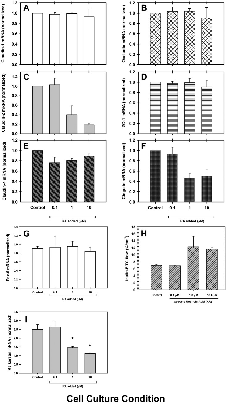
Fig. 8. The disruption of corneal epithelial barrier is in part explained by a decrease in the expression of cldn-2, cldn-4 and cgn.
In RCE1(5T5) epithelial sheets treated with 0.1, 1.0 or 10 µM RA for 48 hours, (F) cgn and (C) cldn-2 showed high susceptibility to RA treatment, while cldn-1 (A), ocln (B) and ZO-1 (D) were not affected. Surprisingly, cldn-4 (E) showed a slight decrease in cultures treated with 0.1 and 1.0 µM, but not with 10 µM RA. At the same time (H), treatment with 1.0 or 10 µM RA for 48 hours induced a 2-fold increase in the paracellular flux of inulin-FITC. (I) Changes in cgn and cldn-2 expression induced by RA were similar to the decrease on the expression of Keratin K3, a late differentiation marker, linked to the expression of corneal terminal phenotype. (G) Given that the expression of the transcription factor Pax-6 did not undergo a significant change after treatment with RA, the results establish a tight link between corneal phenotype expression and TJ assembly. RA seems to modulate phenotype expression of corneal epithelial cells and TJ assembly, whereas Pax-6 seems to regulate corneal epithelial cell commitment. Asterisks indicate statistically significant changes (P≤0.001).

