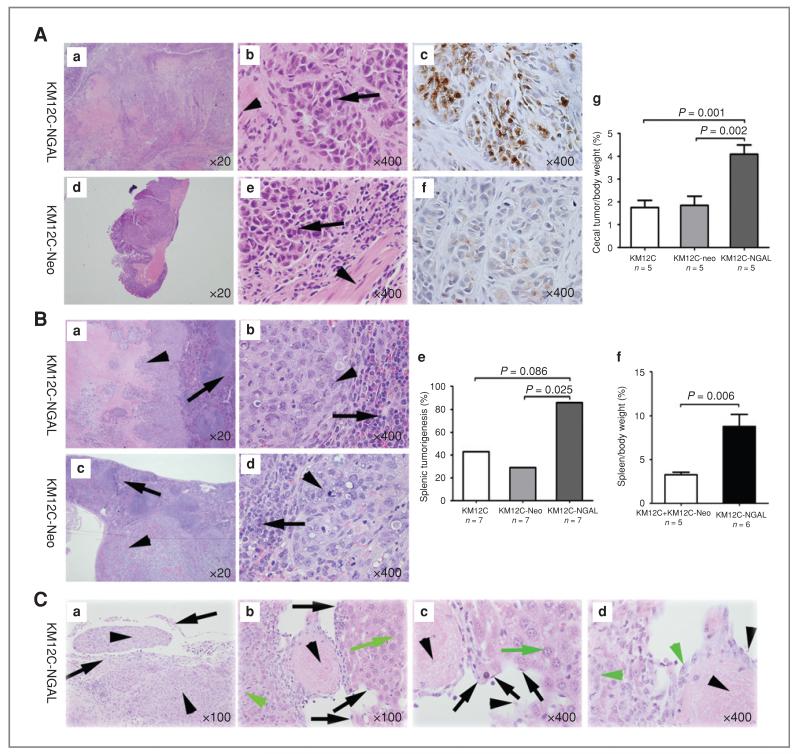
Figure 3.
NGAL overexpression enhanced tumorigenesis and potentially increased liver metastasis of CRC cells in animal models. A, xenografts in cecum after intracecal injection. KM12C-NGAL, KM12C-Neo, or KM12C cells were injected into the cecal wall of nude mice. Tumor cells of the xenografts (arrows) invaded into muscularis (arrowheads) of cecum (b and e). The immunohistochemical staining for NGAL was strong in xenografts of the mice injected with KM12C-NGAL cells (c), and weak in the xenografts of the mice injected with KM12C-Neo cells (f). The percentage of cecal tumor/body weight in the mice injected with KM12C-NGAL was higher than in the mice with KM12C or KM12C-Neo cells (g). B, xenografts in spleen after intrasplenic injection. KM12C-NGAL (a and b), KM12C-Neo (c and d), or KM12C cells were injected into the spleen of nude mice. Arrows denote splenic tissue, and arrowheads indicate tumor cells. The number of mice with splenic xenografts was larger in the group injected with KM12C-NGAL than in the group with KM12C-Neo cells (e). For the mice with splenic tumors, the percentage of spleen/body weight in the mice injected with KM12C-NGAL was higher than that in control groups (f). C, liver metastasis was observed in a nude mouse injected with KM12C-NGAL cells. a, CRC cells (arrowheads) invaded splenic capsule (arrows) in the mouse with liver metastasis; b–d, vascular invasion was found in liver metastasis. Black arrows denote vascular endothelial cells, black arrowheads indicate blood cells, green arrows denote hepatocytes, and green arrowheads indicate metastatic CRC cells.

