Fig. 6.
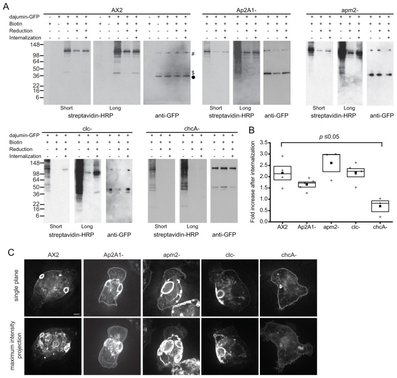
Internalization of dajumin-GFP. (A) Biotin internalization assay for AX2, Ap2A1–, apm2–, clc– and chcA– cells. Dajumin-GFP was immunoprecipitated from cell lysates. Membranes were probed for biotin with streptavidin-HRP (short and long exposures shown) or for GFP with an anti-GFP antibody. Dajumin-GFP full-length protein (#), the 45-kDa fragment ($) and the 40-kDa fragment (•) are indicated. (B) The -fold increase in biotin signal following 15 minutes of internalization calculated from western blots. Solid square, mean; box, s.e.m. and 50th percentile; gray diamond, individual values. Values were compared using a one-way ANOVA. (C) Widefield microscopy images of dajumin-GFP in AX2, Ap2A1–, apm2–, clc– and chcA– cells. A single focal plane (top row) and a maximum intensity projection of the entire cell (bottom row) are shown. Scale bar: 1 µm. See also supplementary material Fig. S2.

