
Fig. 2.
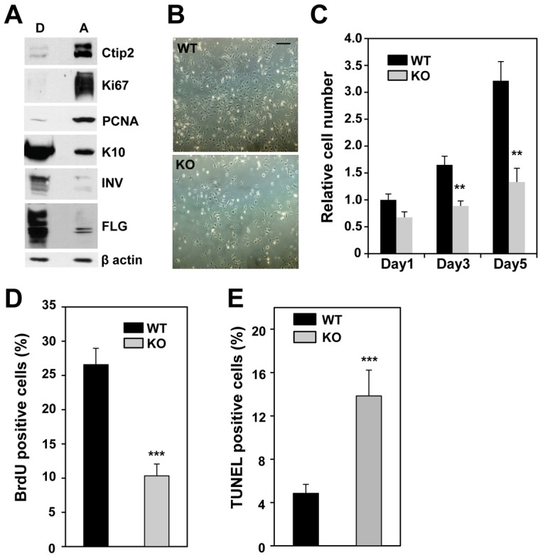
Deletion of Ctip2 results in reduced proliferation and survival of primary mouse keratinocytes in vitro. (A) Cell extracts (10 µg) from attached (A) and detached (D) keratinocytes were immunoblotted with antibodies against Ctip2, Ki67, PCNA, K10, INV, FLG or β-actin (a loading control). (B) Phase-contrast images of primary keratinocytes taken at day 3 after plating. Scale bar: 100 µm. (C) Relative cell numbers were determined using the MTT assay on the indicated days after plating. (D) Subconfluent WT and KO keratinocytes were pulse-labeled with BrdU prior to immunocytochemistry analyses using anti-BrdU antibody and the percentage of BrdU-positive cells was determined. (E) Results of the TUNEL assay for apoptotic cells on fixed, cultured keratinocytes. Statistical significance was determined using Student's t-test (***P<0.001). WT, black bars; KO, gray bars. The results are representative of at least three independent studies.