
Fig. 4.
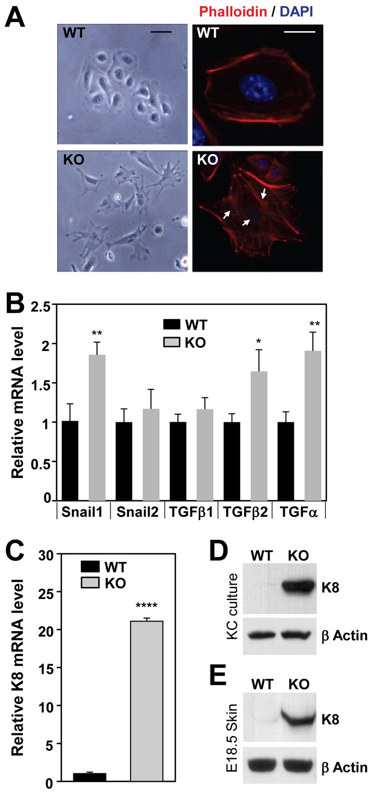
Lack of Ctip2 leads to altered cytoskeletal organization. (A) Phase-contrast images of cultured WT and KO keratinocytes (left panel). Cultured keratinocytes were immunostained with anti-phalloidin (actin staining) and DAPI (nucleus staining) as shown on the right. Scale bar: 20 µm for phase-contrast images; 10 µm for fluorescence image. (B) RT-qPCR analyses of WT or KO primary keratinocytes. Relative mRNA expression levels of the indicated genes were calculated relative to the expression of a housekeeping gene, GAPDH. (C) RT-qPCR analyses of K8 mRNA expression in WT and KO primary keratinocytes (normalized to GAPDH). (D,E) Immunoblotting analyses of cell extracts from WT and Ctip2 KO primary keratinocytes (D) and E18.5 skin (E) using anti-K8 and anti-β actin antibodies. Statistical significance was determined using Student's unpaired t-test (*P<0.05; **P<0.01; ****P<0.0001). The results depicted are representative of at least three independent studies.