Fig. 6.
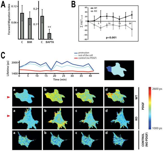
Importance of calcium responses in chemotaxis and generation of Ins(1,4,5)P3 in MEFs treated with PDGF. (A) Forward Migration Index of WT MEFs (immortalised cell population) treated with either PKC inhibitor (BIM) or calcium chelator (BAPTA) was determined using Dunn chambers. The data are from four independent experiments, **P<0.01, ANOVA. (B) Summary of FRET localisation obtained from MEFs expressing Ins(1,4,5)P3 biosensor LIBRA. Fluorescence lifetime imaging (FLIM) was used to monitor conformational changes in LIBRA following Ins(1,4,5)P3 binding that results in an increase in donor lifetime. Plot of the change in difference of FRET index between the membrane regions facing towards (α) and away (β) from the micropipette (Σ FRET α–β) in a number of directionally stimulated MEFs from immortalised cell populations. P<0.001, ANOVA. (C) Illustrations for MEFs expressing LIBRA biosensor (described in B) exposed to PDGF-BB gradient generated by perfusion from a micropipette. Change in the lifetime for a single WT MEF (either in the area of protrusion or in the rest of the cell) over 40 min is shown in the top panel; the area of a single cell or its parts was isolated from a background by applying a mask. The bottom panel shows images of the WT and PLCε KO cells taken before (a, 0 time) and after (b–d, time intervals within a 15 min period) the release of PDGF-BB; non-stimulated WT MEFs were used as a control. Direction of the gradient is shown by red triangles.

