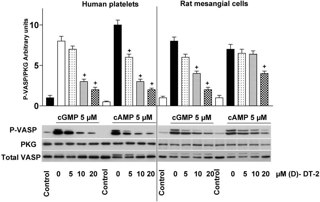
Figure 3.

(D)-DT-2 loses its selectivity against PKG and PKA in cell homogenates. Lysates of human platelets or RMC were pre-incubated (10 min) with indicated concentrations of (D)-DT-2. PKG and PKA were stimulated by cGMP or cAMP, respectively (for 5 min, 5 µM each), Samples were processed for Western blotting. PKG activity was evaluated by VASP-Ser239 phosphorylation (P-VASP). The apparent shift of the VASP signal from 46 kDa to 50 kDa is due to phosphorylation at Ser157. PKG I and total VASP expression are served as loading control. Blots were scanned using SilverFast software and analysed densitometrically by NIH Image J software for uncalibrated optical density. The intensity of P-VASP signal was normalized to the total VASP signal, and then this ratio for each sample was expressed relative to the control which was designated as unity. Results are mean ± SEM, n= 5, +P < 0.05 (Student's t-test). In contrast to in vitro assays (Figure 1), (D)-DT-2 inhibits both PKA and PKG activation in the whole cell lysate.
