
Figure 3.
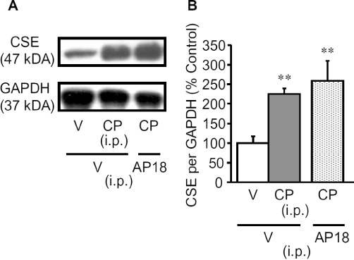
Enhanced expression of CSE protein in the bladder of mice with cyclophosphamide-induced cystitis. (A) Typical photographs of Western blots for CSE in the bladder. (B) CSE protein levels in the bladder quantified by densitometry. The mice were killed for excision of the bladder 4 h after i.p. cyclophosphamide (CP; 300 mg·kg−1) or vehicle. AP18 (10 mg·kg−1) or vehicle (V) was administered i.p. to mice 30 min before cyclophosphamide. Data show the mean with SEM for four to six experiments. **P < 0.01 versus vehicle + vehicle.