
Figure 7.
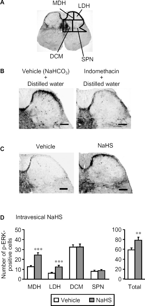
Effect of intravesical NaHS on the number of p-ERK-positive cells in the L6 spinal cord of the mice. The mice were given indomethacin (i.p., 10 mg·kg−1) 55 min before challenge with NaHS. For immunostaining of p-ERK, the spinal cord was transcardially perfused and fixed 5 min after the intravesical administration of NaHS (50 nmol per mouse; 50 µL of 1mM solution). (A) Drawings of four separated regions of the L6 spinal cord in mice. (B) Typical microphotographs for the immunostaining of p-ERK 5 min after distilled water (vehicle) in the mice pretreated with indomethacin or vehicle (NaHCO3). (C) Typical microphotographs for the immunostaining of p-ERK caused by intravesical NaHS in mice pretreated with indomethacin. Scale bars indicate 100 µm. (D) The number of p-ERK-positive cells in the four regions of the bilateral spinal cord following intravesical NaHS. Data indicate the mean with the SEM for 35–40 (seven to eight mice) slices. **P < 0.01, ***P < 0.001 versus vehicle. SPN, sacral parasympathetic nucleus.